PURPOSE OR LEARNING OBJECTIVE:
To investigate the clinical potential of diffusion tensor distribution imaging (DTD) for visually differentiating brain tumors and edema from healthy tissue non-invasively.
METHODS:
Multidimensional diffusion (MDD) MRI images were acquired in 2 lymphoma patients on a 3T Discovery 750w system (GE Healthcare) with a 32-channel head coil. Prototype spin-echo EPI sequences were performed using the following parameters: TR/TE=3298/121 ms, in-plane resolution = 3×3 mm2. MDD consisted of 43 linear and 37 spherical b-tensors at b = 100, 700, 1400, 2000 s/mm2. Total scan time was ~5 min. Post-processing of the data was done using dVIEWR powered by MICE Toolkit (www.dviewr.com). The main features related to average cell density (mean diffusivity, MD) and cell elongation (microscopic anisotropy) can be computed within “bins” corresponding to specific tissue types, i.e., “thin” for elongated cells (e.g., white matter), “thick” for densely packed round cells (e.g., grey matter), “sparse” for low cell-density diffusion environments (e.g., edema) and “big” for free water (e.g., ventricles).
RESULTS:
Bin-resolved segmentation maps (SegM) facilitate the identification of edematous regions, captured by the sparse bin (red areas in SegM). These regions surround the investigated lymphomas, themselves mostly captured by the thin bin (green in SegM), indicating that they consist of elongated cells. These cells are randomly oriented, as they appear white (red+green+blue) in the thin-bin mean-orientation maps (see Figure 1). The bin-resolved MD maps’ colors highlight the inverse relationship between MD and average cell density across different tissue types. In particular, the sparse bin exhibits an intermediate MD characteristic of edema.
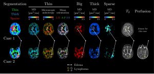
Figure 1. Diffusion Tensor Distribution (DTD) parameter maps of lymphoma cases.
CONCLUSION:
DTD could provide enhanced visualization tools for radiologists aiming to better separate/characterize healthy and pathological tissues non-invasively.
LIMITATIONS:
This pilot study had limitation in terms of small sample size.
